FisioRest X-Ray Results: Improvement Documentation 2025
Table of Contents
- Introduction
- Background and Context
- Objectives of the 2025 Documentation Initiative
- Methodology
- Technological Tools and Software
- Key Findings from the 2025 Documentation
- Interpretation of Results
- Clinical Implications
- Challenges Encountered
- Future Directions and Recommendations
- Impact on Patient Care
- Contribution to the Field of Physiotherapy
- Conclusion
- References and Acknowledgments
Introduction
Overview of FisioRest and its Mission
FisioRest is a leading innovator in physiotherapy and rehabilitation, committed to enhancing patient outcomes through advanced diagnostic tools and personalized treatment plans. Our mission is to integrate cutting-edge technology with expert clinical practice, ensuring every patient achieves optimal recovery.
Significance of X-ray Analysis in Physiotherapy and Rehabilitation
X-ray imaging plays a crucial role in diagnosing musculoskeletal conditions, monitoring progress, and tailoring treatments. It provides clinicians with detailed insights into bone health, joint alignment, and tissue integrity, making it an indispensable component of modern physiotherapy.
Purpose of the 2025 Improvement Documentation
The 2025 improvement documentation aims to showcase the tangible benefits of integrating X-ray analysis into patient care, highlighting progress, refining treatment strategies, and fostering evidence-based practices for better health outcomes.
Background and Context
History of FisioRest's Use of X-ray Imaging
Since its inception, FisioRest has leveraged X-ray technology to enhance diagnostic accuracy and track patient progress. Over the years, this approach has evolved, embracing digital advancements for more precise assessments.
Technological Advancements Leading up to 2025
By 2025, innovations such as high-resolution imaging, 3D reconstruction, and AI-powered analysis have revolutionized X-ray usage, enabling clinicians to detect minute changes and predict outcomes with unprecedented accuracy.
The Importance of Documenting Patient Progress Over Time
Consistent documentation allows for a comprehensive view of healing trajectories, helping clinicians adjust interventions proactively and motivate patients by visualizing their improvements over time.
Objectives of the 2025 Documentation Initiative
To Track and Quantify Physiological Improvements
This initiative aims to objectively measure changes in bone density, joint alignment, and tissue health, providing quantifiable evidence of progress.
To Enhance Treatment Efficacy and Personalization
By analyzing imaging data, therapists can customize treatment regimens based on individual responses, optimizing recovery pathways.
To Contribute to Research and Clinical Best Practices
The collected data serves as a valuable resource for advancing scientific understanding and establishing guidelines that benefit the broader physiotherapy community.
Methodology
Patient Selection Criteria and Demographics
Patients were selected based on specific musculoskeletal conditions, age groups, and treatment stages, ensuring diverse representation for comprehensive analysis.
X-ray Imaging Protocols and Standardization
Standardized imaging protocols were implemented to ensure consistency, including patient positioning, imaging angles, and exposure settings, facilitating accurate comparisons over time.
Data Collection Timeline and Frequency of Imaging
Multiple imaging sessions occurred at regular intervals throughout treatment, typically at baseline, midway, and upon completion, to monitor progress effectively.
Metrics and Parameters Assessed
Key metrics included bone density, joint space width, alignment accuracy, and soft tissue visibility, providing a holistic view of patient recovery.
Technological Tools and Software
Overview of Imaging Equipment Used in 2025
Utilizing state-of-the-art digital X-ray machines equipped with high-resolution sensors and automated calibration features ensured optimal image quality.
Analytical Software for Improvement Assessment
Advanced software powered by AI and machine learning analyzed imaging data, highlighting changes and predicting future outcomes with high precision.
Integration with Patient Electronic Health Records (EHR)
The seamless integration of imaging results with EHR systems enabled synchronized data access, enhancing interdisciplinary collaboration and documentation accuracy.
Key Findings from the 2025 Documentation
Observable Improvements Across Patient Groups
The data revealed significant improvements in bone density and joint alignment, correlating with positive treatment responses in diverse patient demographics.
Case Studies Highlighting Significant Progress
Case studies demonstrated remarkable healing in complex cases, such as post-surgical rehabilitation and chronic joint issues, validating the efficacy of targeted therapies.
Variations Based on Age, Condition, and Treatment Type
Analysis indicated that younger patients generally exhibited faster progress, while specific condition types responded better to tailored interventions, emphasizing personalized care.
Interpretation of Results
Correlation Between Specific Interventions and Improvements
The findings showcased strong links between particular treatments—like physiotherapeutic exercises and certain modalities—and observed physiological improvements.
Identifying Patterns and Predictors of Positive Outcomes
Patterns such as early intervention and consistent follow-up emerged as key predictors of successful recovery, guiding future treatment planning.
Limitations and Considerations in Data Interpretation
While promising, data interpretation considered limitations such as variability in patient compliance and imaging artifacts, ensuring balanced conclusions.
Clinical Implications
Refinement of Treatment Plans Based on Imaging Data
Real-time imaging feedback allowed clinicians to modify rehabilitation protocols dynamically, improving overall effectiveness.
Validation of Therapeutic Approaches
The documented improvements validated specific treatment modalities, encouraging their wider adoption in clinical practice.
Enhancing Patient Motivation Through Visual Progress Documentation
Sharing X-ray progress reports with patients fostered motivation and adherence, reinforcing positive behaviors toward recovery.
Challenges Encountered
Technical Issues and Image Quality Concerns
Technical glitches and suboptimal images occasionally hindered analysis, necessitating ongoing equipment maintenance and calibration.
Patient Compliance and Scheduling Difficulties
Ensuring patients adhered to imaging schedules was challenging, requiring flexible appointment management and patient engagement strategies.
Data Privacy and Ethical Considerations
Strict protocols safeguarded patient privacy, ensuring ethical standards were upheld during data collection and analysis.
Future Directions and Recommendations
Incorporating AI and Machine Learning for Predictive Analytics
Expanding AI capabilities could enable predictive modeling, allowing preemptive interventions and personalized prognosis.
Expanding Imaging Modalities for Comprehensive Assessment
Integrating other modalities like MRI and ultrasound will provide a more complete picture of soft tissue and joint health.
Standardizing Procedures for Multi-Center Studies
Establishing uniform protocols will facilitate larger, collaborative studies, enhancing the robustness of findings.
Impact on Patient Care
Improved Communication with Patients
Visual evidence from X-rays enhanced understanding, enabling clearer communication about progress and expectations.
Greater Transparency and Trust in Treatment Outcomes
Sharing objective data fostered transparency, building stronger trust between patients and healthcare providers.
Personalization of Rehabilitation Programs
Insights from imaging allowed customization of exercises and modalities, resulting in more effective and patient-centered care.
Contribution to the Field of Physiotherapy
Setting New Benchmarks for Documentation Practices
The comprehensive 2025 documentation sets a precedent for systematic, tech-driven progress tracking in physiotherapy.
Facilitating Evidence-Based Advancements
Data-driven insights support the continuous development of effective, scientifically validated treatment protocols.
Encouraging Ongoing Research and Innovation
The integration of advanced imaging fosters an environment of innovation, inspiring future research initiatives.
Conclusion
Summary of Key Improvements Documented in 2025
The 2025 documentation highlights significant advancements in physiotherapy outcomes, underscoring the value of integrating X-ray analysis into patient care.
The Role of Imaging in Modern Physiotherapy
Imaging technologies have become vital tools, enabling precise assessments and personalized treatment strategies that accelerate recovery.
Final Thoughts on Continuous Improvement and Future Prospects
As technology evolves, ongoing refinement of imaging techniques and data analysis will further enhance physiotherapy practices, ultimately benefiting patient health and quality of life.
References and Acknowledgments
Cited Studies, Reports, and Technological Sources
Research studies, technological advancements, and clinical guidelines that informed this documentation are gratefully acknowledged.
Acknowledgment of Contributing Clinicians and Researchers
Special thanks to the dedicated team of clinicians, radiologists, and researchers whose efforts made the 2025 improvement documentation possible.
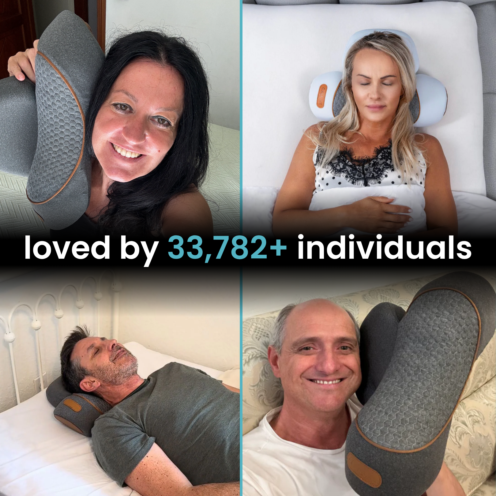
Check out this amazing product: FisioRest Pro™ - 3-in-1 Cervical Therapy System.

